


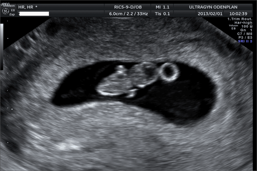


An early ultrasound is typically performed between 6 and 12 weeks of pregnancy. This scan is used to:
- Confirm that the pregnancy is developing (including detecting cardiac activity)
- Verify that the pregnancy is located in the uterus
- Determine the number of embryos
- Estimate gestational age
In some cases, it may be too early to determine gestational age accurately, and a follow-up ultrasound may be recommended.
Why is an early ultrasound done?
An early ultrasound may be recommended for several reasons, including:
- Vaginal bleeding in early pregnancy
- Uncertainty about how far along the pregnancy is
- A history of ectopic pregnancy, to confirm that the pregnancy is located in the uterus
Transvaginal ultrasound in early pregnancy
Before about 10 weeks of pregnancy, a transabdominal ultrasound (on the abdomen) may not provide clear images. In these cases, a transvaginal ultrasound is often used.
During this exam, a small probe is inserted into the vagina to provide clearer images of the uterus and developing pregnancy. It may feel uncomfortable but is generally safe and similar to a pelvic exam.
Light spotting after a transvaginal ultrasound can occur and is usually not harmful.
What does this mean for miscarriage risk?
Seeing cardiac activity on an early ultrasound is associated with a lower risk of miscarriage. As pregnancy progresses, especially after the first trimester, the overall risk decreases further.
If you have experienced a previous miscarriage or complications, your healthcare provider may recommend an early ultrasound for reassurance and monitoring.
Sources: