


Most pregnant people in the US are offered a detailed ultrasound during the second trimester. This is often called the anatomy scan and is usually performed between 18–22 weeks of pregnancy.
The purpose of the scan is to:
- check how the baby is developing
- confirm how many babies you are carrying
- look at the position of the placenta
If your due date has not already been confirmed earlier in pregnancy, measurements from the ultrasound may also be used to estimate gestational age.
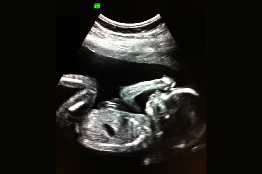
During the scan, the sonographer or healthcare provider carefully examines the baby’s anatomy using ultrasound. This includes the brain, spine, heart, stomach, kidneys, arms and legs. While many structural conditions can be identified, not all abnormalities or medical conditions can be detected during an anatomy scan.
Before your anatomy scan
- Some clinics may ask you to arrive with a partially full bladder, as this can help improve the images.
- You are usually welcome to bring your partner or another support person.
- The scan typically takes around 30–45 minutes.
- You will lie on an exam table while gel is applied to your abdomen to create clearer ultrasound images.
- The examination is painless, although slight pressure on the abdomen can sometimes feel uncomfortable.
Sources: